1752119111@qq.com
+86 1365 2921 391
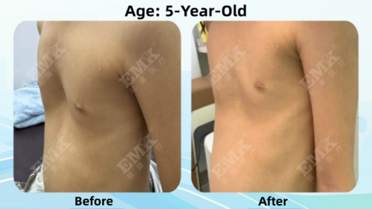
5-Year-Old Child Pectus Excavatum Case: Before and After 2 Years of Vacuum Bell Treatment
Pectus Excavatum is one of the most common chest wall deformities in children and is characterized by a sunken sternum and inward depression of the chest wall.
At around five years of age, chest wall development becomes more noticeable, and some parents begin to recognize differences in chest shape during daily activities, sports, or swimming.
This case presents the chest wall appearance of a 5-year-old child before treatment and after 2 years of Vacuum Bell therapy.
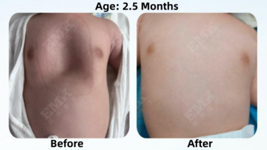
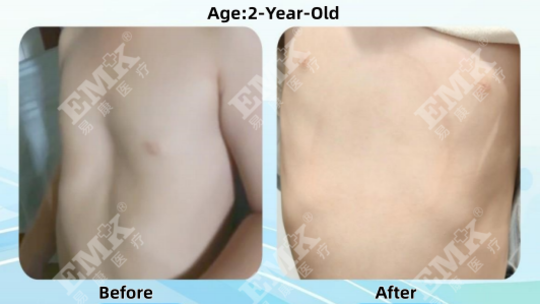
5 years old
July 2024
Visible central chest depression
Uneven anterior chest wall contour
Typical funnel-shaped chest appearance
Chest shape difference noticed by parents
Vacuum Bell Therapy
Non-surgical chest wall management
Improve chest appearance
Reduce visible depression
Improve chest symmetry
Support normal chest wall development
A noticeable depression of the sternum was present, creating a visible funnel-shaped chest appearance.
Compared with baseline:
Reduced sternal depression
Improved chest wall contour
More natural chest appearance
Better chest symmetry
Based on image comparison:
Improved compared with the initial assessment.
Smoother anterior chest wall appearance.
More balanced chest wall structure.
Closer to a natural chest shape.
Around age five, the chest wall continues to grow and develop.
Regular evaluation may help:
Monitor chest wall development
Track progression of the deformity
Support long-term management planning
Guide future treatment decisions
Yes. In some cases, the deformity becomes more apparent during childhood and adolescence.
Suitability depends on chest wall flexibility, severity of the deformity, and professional evaluation.
Yes. Regular monitoring can help assess chest wall growth and development.
The chest wall remains relatively flexible during early childhood.
Through regular follow-up and individualized management, chest wall development and chest contour changes can be monitored over time.
Treatment planning should always be tailored to the patient's anatomy and growth pattern.
This case is provided for educational purposes only.
Individual outcomes vary depending on anatomy, severity, growth patterns, and treatment response.
The results shown in this case do not guarantee similar outcomes for all patients.
EMK Yikang Medical provides:
✓ Professional Chest Wall Evaluation
✓ CT Imaging Analysis
✓ 3D Chest Wall Reconstruction
✓ Customized Design Based on CT or 3D Chest Modeling
✓ Long-Term Follow-Up Support
Contact our team today for a personalized assessment.
Contact: KAM
Phone: +86 1365 2921 391
Tel: +86 1365 2921 391
Email: 1752119111@qq.com
Add: Orthosis Customization Center, 6th Floor, Rehabilitation Building, Guangdong Maternal and Child Health Hospital
We chat